Section
10:
Wound Complications -
Fungating
Wounds
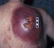
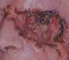
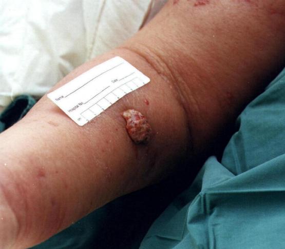
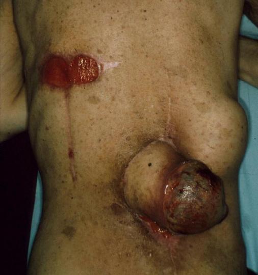
Fungating wounds are caused either by a local tumour infiltrating the skin, or by metastatic spread from the primary tumour. Malignant fungating lesions are an immense challenge. Management goals vary depending on the stage of the underlying cancer, the patient’s prognosis, and the individual’s own goals and wishes. In some cases, the aim is to arrest tumour growth, but in many situations fungating tumours occur at the end of life, and treatment is completed in a palliative setting that focuses on comfort and maintenance of the best possible quality of life for that patient and their family. In either case it is important to remember that the symptoms produced by a fungating wound are often as distressing as the wound itself. Management focuses on alleviation of distressing symptoms including pain, cutaneous irritation, exudate, bleeding and odour.
Treatment options
Wound exudate is produced as a normal part of the wound healing process.
In fungating wounds the exudate produced can be excessive. This is thought to be due to increased permeability of blood vessels within the tumour and secretion of vascular permeability factor by tumour cells.
-
Remove slough/necrotic debris
-
Manage excess exudate
-
Keep moist
Heavy
exudate
Fibrous Hydrocolloid
Alginate
Extra-absorbent Foam
Light exudate
Hydrogel
Fibrous Hydrocolloid
Non-adhesive Foam

Blood vessels can be disrupted by the infiltration of tumour cells which can lead to bleeding at the wound site.
Traumatic dressing changes can also lead to bleeding.
-
Control bleeding
- Prevent trauma at dressing change
A Silicone Wound Contact Layer should be used as a primary dressing to prevent adherence to the wound.
Malodour
Malodour is associated with necrotic tissue that supports the growth of anaerobic bacteria, and the presence of volatile fatty acids in the wound. Stagnant exudate, infection and fistula formation are also contributing factors.
Malodour is reported to be the most distressing symptom from the patients perspective.
-
Reduce odour
-
Check for infection via swab
Metronidazole gel
- apply to clean wound 1-2 times daily and cover with non-adherent dressing.
CarboFlex® should be used for moderately exuding wounds and CliniSorb® for lightly exuding wounds.
Skin
irritation
-
Protect from maceration/breakdown
-
Control itching
Cavilon®, Diprobase® or Dermol® may provide relief.
Zinc oxide paste can be used to prevent peri-wound maceration.
A Hydrogel Sheet can also reduce itching and promote comfort.
There are several types of pain associated with malignant wounds: deep pain, neuropathic pain and superficial pain related to procedures.
Dressing removal has been found to be the time of greatest pain for those with chronic wounds.
- Analgesic drugs should be prescribed using the World Health Organisation (WHO) guidelines for the control of cancer pain and in accordance with the Tayside Area Formulary (TAF) - CLICK HERE
A short-acting analgesic such as Oramorph® may be given 20-30 minutes prior to dressing changes.
Where nerve pain exists, adjuvant therapies may be of benefit. Please see TAF section 4.7.3 for more information.
The use of morphine or diamorphine mixed with a hydrogel (to form a 0.08–0.1% mixture) and applied topically to the wound has been found to be an effective method of pain relief in ulcerating wounds, including fungating wounds.